It is suggested that high quality MRI is performed first when surgical evaluation is undertaken and if negative the patient carefully counselled before proceeding with any investigations, as successful resective surgery is an unlikely outcome in such MRI negative cases.īetween January 1995 and January 1997, 222 patients attending the National Hospital for Neurology and Neurosurgery epilepsy clinics had video-EEG telemetry as part of their investigation for epilepsy surgery.Īll patients had high resolution MRI performed using a 1.5 Tesla GE Signa machine, with a protocol for T1 weighted coronal images in thin contiguous slices 1.5 mm or less and T2 weighted images as described previously. In five of the 40, evaluation led to a hypothesis that could be tested by intracranial studies three proceeded to surgery. None of 40 disclosed a well localised epileptogenic zone concordant with other tests that would have allowed the patient to proceed directly to surgery. All patients undergoing presurgical evaluation at the National Hospital for Neurology and Neurosurgery between 19 were reviewed and 40 were identified without definite MRI abnormalities.

The aim was to consider whether these patients were likely to proceed to surgical treatment after scalp video-EEG telemetry. Be sure to speak up if there’s anything about your results that you don’t understand.When considering surgery for intractable partial seizures, even with high resolution MRI, some patients do not show structural abnormalities. Before you review the results, it may be helpful to write down any questions you might want to ask. It’s very important to discuss your test results with your doctor.


A technician will measure your head and mark where to place the electrodes.You’ll lie down on your back in a reclining chair or on a bed.The test usually takes roughly 30 to 60 minutes to complete and involves the following steps: Specialized technicians administer EEGs at hospitals, doctor’s offices, and laboratories. The electrodes transfer information from your brain to a machine that measures and records the data. An electrode is a conductor through which an electric current enters or leaves. Other factors that can influence your EEG reading include:Īn EEG measures the electrical impulses in your brain by using several electrodes attached to your scalp. The person responsible for interpreting your EEG will take these movements into account. Several types of movements can potentially cause “artifacts” on an EEG recording that mimic brain waves. Factors that could interfere with an EEG reading Some people may not be able to hyperventilate safely, such as people with a history of stroke, asthma, or sickle cell anemia. Hyperventilation is also commonly induced during an EEG to produce abnormalities.

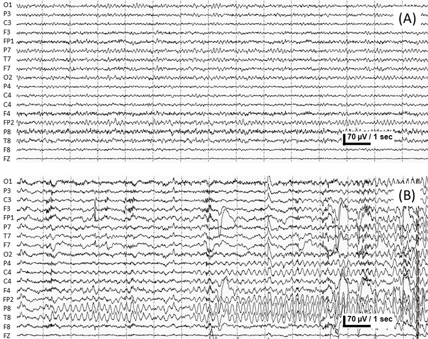
The technician performing the EEG is trained to safely manage any situation that might occur. When someone has epilepsy or another seizure disorder, there’s a small risk that the stimuli presented during the test (such as a flashing light) may cause a seizure. If an EEG does not produce any abnormalities, stimuli such as strobe lights, or rapid breathing may be added to help induce any abnormalities.


 0 kommentar(er)
